Lokavarapu Manoj Joshua1, Farhanul Huda1, Shalinee Rao2, Bina Ravi1
1Department of Surgery, All India Institute of Medical Sciences, Rishikesh, Uttarakhand, India
2Department of Pathology, All India Institute of Medical Sciences, Rishikesh, Uttarakhand, India
| Date of Submission | 02-Apr-2020 |
| Date of Decision | 17-Jun-2020 |
| Date of Acceptance | 30-Aug-2020 |
| Date of Web Publication | 05-Dec-2020 |
Correspondence Address:
Lokavarapu Manoj Joshua
Room 230, Block 86, Department of Surgery, All India Institute of Medical Sciences, Rishikesh, Uttarakhand
India
Source of Support: None, Conflict of Interest: None
DOI: 10.4103/jcar.JCar_9_20
Abstract
BACKGROUND: Filamin A is an actin-crosslinking protein expressed in many malignancies, although its prognostic and therapeutic role in breast cancer is not studied. There is enigma regarding its dual role in cancer, the tumor-progressing or tumor-suppressing effects depending on the site to which it localizes in the cell. The current study aimed to detect Filamin A expression in breast cancer and its association with other biomarkers and other clinicopathological parameters and established risk factors in breast cancer so that it can be a potential site for targeted therapy.
MATERIALS AND METHODS: One hundred female patients of histologically proven breast cancer who presented to our hospital over a 2-year period were included in the study. None of the patients received prior radiotherapy, chemotherapy, or immunotherapy. Patients with recurrent breast cancer are not included in the study. All study cases are subjected to immunohistochemistry for estrogen receptor, progesterone receptor, Her2 neu, and ki-67 from core biopsy tissue of cases diagnosed as breast carcinoma. Tissue sections were subjected to immunohistochemistry with anti-Filamin A.
RESULTS: Filamin A is expressed in 69% of cases of invasive breast cancer in our study. There was no statistically significant relationship of Filamin A immunoexpression with histological grade, age, parity, oral contraceptive use, smokeless tobacco use, TNM staging, clinical staging, clinical prognostic staging, and also ER, PR, Her2 neu, and ki-67 status (P > 0.05). Thus, it appears to be an independent biomarker in breast carcinoma. Filamin A was expressed only in the cytoplasm in all our study cases. Filamin A expression can be observed in adjacent normal breast tissue and benign fibroadenoma tissues also, but the pattern of expression is mainly membranous with cytoplasmic positivity. The cytoplasmic expression is seen in malignant cells as well as normal breast and benign tumor sections implicating the dual role of Filamin A in breast cancer.
CONCLUSION: No significant correlation could be found between Filamin A expression and clinicopathological parameters in our study. The cytoplasmic expression is seen in malignant cells as well as normal breast and benign tumor sections implicating the dual role of Filamin A in breast cancer. Filamin A immunoexpression should be further correlated with metastasis-free survival period of breast cancer patients
Keywords: Breast cancer, Filamin A, immunohistochemistry, risk factors
| How to cite this article: Joshua LM, Huda F, Rao S, Ravi B. Clinicopathological significance of immunohistochemical expression of Filamin A in breast cancer. J Carcinog 2020;19:13 |
| How to cite this URL: Joshua LM, Huda F, Rao S, Ravi B. Clinicopathological significance of immunohistochemical expression of Filamin A in breast cancer. J Carcinog [serial online] 2020 [cited 2021 Oct 13];19:13. Available from: https://carcinogenesis.com/text.asp?2020/19/1/13/302494 |
Introduction
Breast cancer is the second most common malignancy accounting for 11.6% of all new cases diagnosed and 6.6% of all cancer-related deaths, next only to lung cancer worldwide.[1] Investigation of novel biomarkers leads to better understanding of tumor biology and the clinical utility of existing biomarkers. Filamin A is a cytoskeletal protein which crosslinks actin into orthogonal networks. The actin cytoskeleton plays a role in cell division, cell shape, motility, and signal transduction.[2],[3] FLNa expression may contribute to tumorigenesis. FLNa overexpression is associated with metastasis in lung cancer.[4] In melanoma, FLNa positive tumor cells are found to be more invasive than FLNa negative cells.[5] Its prognostic and therapeutic role in breast cancer is not studied. There is enigma regarding its dual role in cancer, the tumor-progressing or tumor-suppressing effects depending on the site to which it localizes in the cell. The current study aimed to detect Filamin A expression in breast cancer and its association with other biomarkers and other clinicopathological parameters and established risk factors in breast cancer so that it can be a potential site for targeted therapy.
Materials and Methods
One hundred female patients of histologically proven breast cancer who presented to our hospital over a 2-year period were included in the study. None of the patients received prior radiotherapy, chemotherapy, or immunotherapy. Patients with recurrent breast cancer are not included in the study. Clinical TNM staging was done according to the AJCC 8th edition 2017. All study cases are subjected to immunohistochemistry for estrogen receptor, progesterone receptor, Her2 neu, and ki-67 from core biopsy tissue of cases diagnosed as breast carcinoma. Four-micron thick sections are cut from paraffin-embedded tissue blocks and were subjected to immunohistochemistry with anti-Filamin A/FLNa Picoband rabbit IgG polyclonal antibody (Boster Biological Technology, Pleasanton, CA, USA) in lyophilized form at dilution of 0.5 mg/ml with antibody diluent.
Sections were examined under low-power (×100) and high-power (×200 and ×400) magnification to observe immunoreactivity. Sections of benign skin from the chest wall of female patients, distant normal breast tissue, and sections from benign fibroadenoma are used as negative control for anti-Filamin A antibody. Criteria for immunopositivity are membrane and cytoplasmic positivity of malignant cells. Immunopositivity is scored from 0 to 3+ (Filamin score); the intensity of immunopositivity was scored as zero if no staining it is scored as 1+ for weak cytoplasmic staining in <10% of cells 2+ for moderate cytoplasmic staining in >10% of cells and 3+ for marked cytoplasmic staining in >10% of the cells. A score of 0 or 1 was considered as negative result for FLNa expression (low), whereas scores of 2+ or 3+ were considered as positive (high) FLNa expression. Immunoexpression of FLNa protein was correlated with clinical stage, clinical prognostic stage, and histological grade of tumor. Immunoexpression of FLNa protein will be correlated with parameters such as age, age at first child, menstruation status, use of oral contraceptive pills and hormone replacement therapy, nulliparity, tobacco use, alcohol use, and family history of breast cancer. Statistical analysis was done using SPSS version 23. T-test, Chi-squared test, Fisher’s exact test, Wilcoxon test, and Kruskal–Wallis test were used to test significance.
Statistical analysis
Filamin A was expressed only in cytoplasm in all our study cases. Filamin A expression can be observed in adjacent normal breast tissue and benign fibroadenoma tissues also, but pattern of expression is mainly membranous with cytoplasmic positivity. There was no statistically significant association of immunoexpression of Filamin A and with the age (P = 0.958), parity (P = 0.219), oral contraceptive usage (P = 1.0), smokeless tobacco usage (P = 1.0) of patients. The immunoexpression of Filamin A was not associated with histological grade (P = 0.072), T stage (P = 0.812), N stage (P = 0.644), M staging (P = 0.141), clinical staging (P = 0.386), clinical prognostic staging (P = 0.678), and also ER (P = 0.241), PR (P = 0.643), Her2 neu (P = 0.582), ki-67 status (P = 0.181) of tumor [Figure 1].
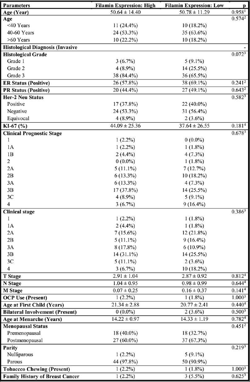 |
Figure 1: Association of Filamin A expression with clinicopathological parameters Click here to view |
Discussion
Filamin A has been studied in a number of cancers such as prostate and lung cancer. The possible role of its expression and localization in the malignant cell has not been established in breast cancer. In one study, Filamin A, it is shown to suppress breast cancer cell migration and invasion.[2],[6] In another study, it was shown that the silencing of Filamin A inhibited migration and invasion by cancer cells[7] We studied its expression and possible correlation with other established biomarkers and risk factors of breast cancer in this study. In our study, age majority of the patients are between 40 and 60 years [Figure 1]. Thirty-six percent of the patients are premenopausal and 64% of the patients are postmenopausal. The median age at the first child in our study is 20 years, which is similar to current demographic trends in India.[8] Only 2% of the patients have a history of oral contraceptive usage and none of the patients has a history of hormone replacement therapy. In our present study, none of the patients had a history of smoking or alcohol consumption. About 2% of the patients had smokeless tobacco use (tobacco chewing). The role of smokeless tobacco in breast cancer is investigated in very few studies.
Pattern of expression
Filamin A is expressed in 69% of cases of invasive breast cancer in our study. Tian et al. reported 63.54% and Guo et al. reported 52.6% positivity in their studies; a slightly different scoring system was used in the study done by Guo et al.[9],[10] Studies have shown that when Filamin A localizes to the cytoplasm, it has a tumor-promoting effect and when it localizes to the nucleus, it has tumor-suppressing effect.[11]
Filamin A was expressed only in the cytoplasm in all our study cases. Filamin A expression can be observed in adjacent normal breast tissue and benign fibroadenoma tissues also, but the pattern of expression is mainly membranous with cytoplasmic positivity [Figure 2]. The intensity of immunopositivity is scored from 0 – 3+ [Figure 3],[Figure 4],[Figure 5],[Figure 6]. Tian et al. have observed cytoplasmic observation mainly at the edge of cells and intercellular substance in malignant cells and no immunoreactivity for Filamin A in normal breast tissue and benign tumor sections.[9] In the study done by Guo et al., immunoreactivity was observed in normal breast tissue.[10] In a study done by Jiang et al., Filamin A expression was noted in the cytoplasm.[12] In another study, Filamin A was observed to be expressed within the cytoplasm and cell membrane.[13] Previous studies failed to find an association between localization of Filamin A and breast cancer.
| Figure 2: Left: Photomicrograph showing Filamin A expressivity in nonneoplastic breast tissue showing membranous and cytoplasmic positivity (×10). Right: Photomicrograph showing a membranous pattern in myoepithelial cells in fibroadenoma (×10) Click here to view |
 |
Figure 3: Left: Photomicrograph showing invasive carcinoma of the breast (H and E, ×40). Right: Immunohistochemistry: Score 0 (absent) (×40) photomicrograph showing the absence of Filamin A expression in malignant cells Click here to view |
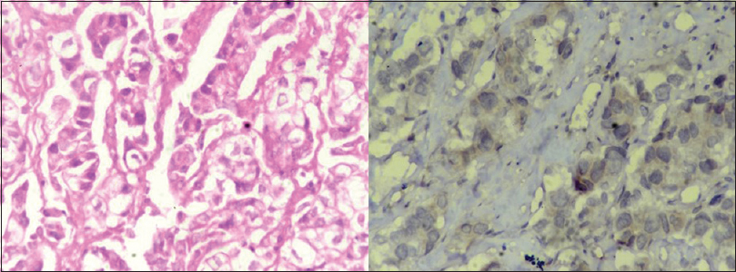 |
Figure 4: Left: Photomicrograph showing invasive carcinoma of the breast (H and E, ×40). Right: Immunohistochemistry: Score 1 (mild positivity) (×40); photomicrograph showing mild cytoplasmic positivity of Filamin A expression in malignant cells Click here to view |
| Figure 5: Left: Photomicrograph showing invasive carcinoma of the breast (H and E, ×40). Right: Immunohistochemistry: Score 2 (moderate positivity) (×40); photomicrograph showing moderate cytoplasmic positivity of Filamin A expression in malignant cells Click here to view |
| Figure 6: Left: Photomicrograph showing invasive carcinoma of the breast (H and E, ×40). Right: Immunohistochemistry: Score 3 (strong positivity) (×40); photomicrograph showing strong cytoplasmic positivity of Filamin A expression in malignant cells Click here to view |
Association between Filamin A expression and clinicopathological parameters
No significant association was identified between the age of patient and Filamin A expression (P = 0.574), which was consistent with the other studies [Figure 1].[9],[10] High Filamin A expression was associated with higher histological grade, but there was no statistically significant relationship between Filamin A expression and histological grade of tumor (P = 0.072). The ER, PR, and Her2 neu status was not shown to have a significant association with Filamin A expression.
In a study done by Guo et al., positive association has been observed between PR status and Filamin A expression, whereas in the study done by Tian et al., there was no significant relationship between hormone receptor status and Filamin A expression.[9],[10] In our study, there was no significant relationship between the TNM stage, clinical stage, clinical prognostic stage, lymph node metastases, and Filamin A expression (P > 0.05) [Figure 1]. In the study done by Tian et al., Filamin A expression was shown to have a significant association with the TNM stage, lymph node metastasis, and vascular and perineural invasion, whereas in the study done by Guo et al., there was no association with lymph node metastasis.[9]
In our study, we studied the association of ki-67, a proliferation marker with Filamin A score and Filamin A expression which was not previously studied and found to be statistically insignificant. Previous studies have shown that Filamin A modulates chemosensitivity in triple-negative breast cancer.[14] There was no significant relationship between Filamin A expression and age at menarche, menopausal status, usage of oral contraceptive pills, and parity. Menstruation status has shown to be significantly associated with Filamin A expression (P = 0.038) in other studies.[9]
Conclusion
No significant correlation could be found between Filamin A expression and clinicopathological parameters in our study. The cytoplasmic expression is seen in malignant cells as well as normal breast and benign tumor sections implicating the dual role of Filamin A in breast cancer. Filamin A immunoexpression should be further correlated with metastasis-free survival period of breast cancer patients.
Financial support and sponsorship
Nil.
Conflicts of interest
There are no conflicts of interest.
References
| 1. |
Bray F, Ferlay J, Soerjomataram I, Siegel RL, Torre LA, Jemal A. Global cancer statistics 2018: GLOBOCAN estimates of incidence and mortality worldwide for 36 cancers in 185 countries. CA Cancer J Clin 2018;68:394-424.
 |
| 2. |
Feng Y, Walsh CA. The many faces of filamin: A versatile molecular scaffold for cell motility and signalling. Nat Cell Biol 2004;6:1034-8.
|
| 3. |
Popowicz GM, Schleicher M, Noegel AA, Holak TA. Filamins: Promiscuous organizers of the cytoskeleton. Trends Biochem Sci 2006;31:411-9.
|
| 4. |
Uramoto H, Akyürek LM, Hanagiri T. Positive relationship between filamin and VEGF in patients with lung cancer. Anticancer Res 2010;30:3939-44.
|
| 5. |
Flanagan LA, Chou J, Falet H, Neujahr R, Hartwig JH, Stossel TP. Filamin A, the Arp2/3 complex, and the morphology and function of cortical actin filaments in human melanoma cells. J Cell Biol 2001;155:511-7.
|
| 6. |
Xu Y, Bismar TA, Su J, Xu B, Kristiansen G, Varga Z, et al. Filamin A regulates focal adhesion disassembly and suppresses breast cancer cell migration and invasion. J Exp Med 2010;207:2421-37.
|
| 7. |
Ji ZM, Yang LL, Ni J, Xu SP, Yang C, Duan P, et al. Silencing filamin A inhibits the invasion and migration of breast cancer cells by up-regulating 14-3-3s. Curr Med Sci 2018;38:461-6.
|
| 8. |
India – Median Age at First Pregnancy by Age Groups 2014 | Statista. Available from: https://www.statista.com/statistics/680256/median-age-at-first-pregnancy-by-age-groups-india/. [Last accessed on 2019 Oct 26].
|
| 9. |
Tian HM, Liu XH, Han W, Zhao LL, Yuan B, Yuan CJ. Differential expression of filamin A and its clinical significance in breast cancer. Oncol Lett 2013;6:681-6.
|
| 10. |
Guo Y, Li M, Bai G, Li X, Sun Z, Yang J, et al. Filamin A inhibits tumor progression through regulating BRCA1 expression in human breast cancer. Oncol Lett 2018;16:6261-6.
|
| 11. |
Shao QQ, Zhang TP, Zhao WJ, Liu ZW, You L, Zhou L, et al. Filamin A: Insights into its exact role in cancers. Pathol Oncol Res 2016;22:245-52.
|
| 12. |
Jiang X, Yue J, Lu H, Campbell N, Yang Q, Lan S, et al. Inhibition of filamin-A reduces cancer metastatic potential. Int J Biol Sci 2013;9:67-77.
|
| 13. |
Zhong Z, Yeow WS, Zou C, Wassell R, Wang C, Pestell RG, et al. Cyclin D1/cyclin-dependent kinase 4 interacts with filamin A and affects the migration and invasion potential of breast cancer cells. Cancer Res 2010;70:2105-14.
|
| 14. |
Zhao P, Ma W, Hu Z, Zang L, Tian Z, Zhang K. Filamin A (FLNA) modulates chemosensitivity to docetaxel in triple-negative breast cancer through the MAPK/ERK pathway. Tumour Biol 2016;37:5107-15.
|